Real-time analysis for medical diagnostics

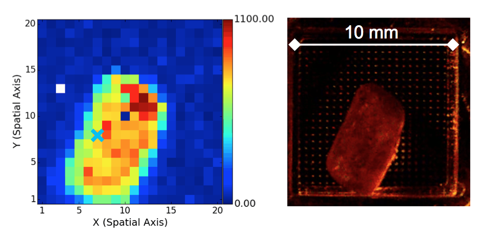
The images show a camera shot and the corresponding Raman image, i.e. the spatial dispersion of sucrose. Experts recognise the false colours as the intensity of the CH2 torsional vibration signal at 850 cm-1.
In the context of current technology transfer projects, scientists at AIP have managed to successfully apply the spectral imaging method, developed in astrophysics, to diagnostics in the field of medicine. In contrast to digital cameras, which only register a brightness value for each pixel, this method detects an entire spectrum. AIP has made a name for itself internationally with this method, referred to also as integral field spectroscopy (IFS). The method is used for instruments such as PMAS and MUSE.
In a recent publication, researchers led by Elmar Schmälzlin have shown for the first time that medical imaging based on IFS produces not only single images, but can be used to record whole image sequences, i.e. videos.
The responsible project manager Martin Roth stated: “Our team has made a significant breakthrough in this field: for the first time, medical practitioners have the prospect of a real-time minimally invasive optical system for diagnostics that surgeons can use to carry out the biopsy and complete removal of potentially cancerous tissue in one single step in the future.”
Recent scientific evidence has shown that the determination of resection borders, i.e. the differentiation between healthy and cancerous tissue, can also take place without a previous laboratory assessment by the pathologist. Resection borders can now be determined directly on the patient using a fibre probe and the method of Raman spectroscopy. This method involves the evaluation of what is referred to as a ‘spectral fingerprint’, which is characteristic for the different types of tissue, similar to the method used by astrophysicists to measure the age and chemical composition of stars and gaseous nebulae. Roth added: “Today’s commercially available spectrographs can just about produce a spectrum for a single measuring point. Unlike astronomers, who can take their time to carefully study spectra sitting at the computer, surgeons in the process of undertaking operations require reliable information about the relevant tissue in the shortest time possible, i.e. a complete picture, preferably in real time.”
There is still a long way to go until this can be achieved. Scientists at AIP are collaborating with medical scientists from the Department of Dermatology, Venerology and Allergology at the Charité-Universitätsmedizin in Berlin to validate the method in order to demonstrate the reliability of imaging Raman spectroscopy – an important milestone on the path towards clinical trials involving an optimised device. Preliminary tests conducted at AIP on the principal feasibility of a future video Raman method are promising.
AIP researchers initially tested video Raman technology in a series of laboratory tests. A lump of sugar dissolving in water was used as the model system. The dissolution process was documented by video Raman. It took ten seconds to record a single image, followed by ten seconds readout time, resulting in an effectively achieved frame rate of twenty seconds. The time limitation was mainly caused by the technical properties of the detector chip used. Scientists are currently working on a faster camera system, which should enable the user-specific selection of the read-out area.
As a member of the Leibniz Research Alliance “Medical Technology”, AIP is committed to the transfer of knowledge and technology from astrophysics to medicine www.lfv-medizintechnik.de/.
This research is funded by the German Federal Ministry of Education and Research under the programmes VIP (03V0843), ZIK (03Z2AS1) and Zwanzig20 (03ZZ0423).
innoFSPEC Potsdam is a joint initiative of the Leibniz Institute for Astrophysics Potsdam (AIP) and the Physical Chemistry Group at the University of Potsdam. Their work focuses on basic research and the development of innovative technological solutions for fibre sensing and multi-channel spectroscopy. The centre for innovation competence combines the skills of new methods of chemical analysis with high-performance multi-object and multi-channel spectroscopy. In the process, it bases its methods on rapid developments in the field of photonics. In addition, innoFSPEC Potsdam is actively involved in promoting technology transfer, teaching and networking, and undertakes cooperative activities with regional and international industrial enterprises and research institutions.
Science contact: Prof. Dr. Martin M. Roth, mmroth@aip.de, +49 331-7499 313
Media contact: Kerstin Mork, presse@aip.de, +49 331-7499 803
Further information
Publication "The Potsdam MRS Spectrograph - heritage of MUSE and the impact of cross-innovation in the process of technology transfer" on arXiv.org
Publication "Ultrafast imaging Raman spectroscopy of large-area samples without stepwise scanning" in Journal of Sensors and Sensor Systems
AIP press release "Cancer Diagnostics with Astrophysics", 14 October 2013
Images
The images show a camera shot and the corresponding Raman image, i.e. the spatial dispersion of sucrose. Experts recognise the false colours as the intensity of the CH2 torsional vibration signal at 850 cm-1.